Call +(254) 703 030 000 / 751 483 999 / 721 704 777
.....Read More
Frequently Asked Questions
What is intubation and why is it necessary?
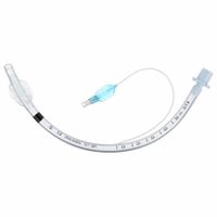
Intubation is a medical procedure involving the insertion of a tube into the trachea (windpipe) to maintain an open airway, facilitate ventilation, or administer certain medications. This procedure is typically performed in emergency situations, during surgery, or when a patient is unable to breathe independently.
The necessity of intubation arises in several scenarios:
1. **Airway Protection**: In cases of trauma, neurological impairment, or decreased consciousness, the risk of airway obstruction increases. Intubation ensures the airway remains open and protected from aspiration of gastric contents or foreign materials.
2. **Respiratory Failure**: Conditions like severe asthma, chronic obstructive pulmonary disease (COPD), pneumonia, or acute respiratory distress syndrome (ARDS) can lead to inadequate oxygenation or ventilation. Intubation allows for mechanical ventilation, providing the necessary support for gas exchange.
3. **Surgical Procedures**: During general anesthesia, intubation is often required to secure the airway and facilitate controlled ventilation, ensuring the patient receives adequate oxygen and anesthetic gases.
4. **Severe Infections**: Infections such as sepsis can lead to respiratory compromise. Intubation may be necessary to support breathing and manage the increased metabolic demands.
5. **Cardiac Arrest**: In cases of cardiac arrest, intubation can be crucial for effective cardiopulmonary resuscitation (CPR), ensuring oxygen delivery to vital organs.
Overall, intubation is a critical intervention in managing airway emergencies, supporting respiratory function, and ensuring patient safety during medical procedures.
How does suction equipment help in airway management?
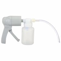
Suction equipment plays a crucial role in airway management by removing obstructions that can impede breathing. It is used to clear secretions, blood, vomit, or other fluids from the airway, ensuring that the patient maintains a clear passage for air exchange. This is particularly important in emergency situations, during surgical procedures, or in patients with compromised airway reflexes.
The equipment typically consists of a suction pump, a collection canister, and a suction catheter. The suction pump generates negative pressure, which helps in aspirating fluids from the airway. The catheter, which is inserted into the patient's mouth, nose, or tracheostomy tube, is flexible and designed to reach the obstruction without causing trauma to the airway tissues.
In emergency settings, such as cardiac arrest or trauma, rapid suctioning can prevent aspiration and improve ventilation. In surgical settings, it helps maintain a clear field for intubation and extubation. For patients with chronic conditions, such as those with neuromuscular disorders, suctioning is essential for routine airway clearance.
Suction equipment also aids in preventing complications like hypoxia, aspiration pneumonia, and atelectasis by ensuring that the airway remains unobstructed. It is a vital component of airway management protocols in hospitals, ambulances, and home care settings, providing both immediate and ongoing support for patients with airway management needs.
What are the risks associated with intubation?
Intubation, while a common and often necessary medical procedure, carries several risks:
1. **Trauma and Injury**: Intubation can cause trauma to the teeth, lips, tongue, vocal cords, and trachea. Dental damage is particularly common.
2. **Aspiration**: There is a risk of stomach contents entering the lungs, leading to aspiration pneumonia, especially if the patient has not fasted.
3. **Hypoxia**: Delays or difficulties in intubation can lead to inadequate oxygenation, resulting in hypoxia, which can cause brain damage or cardiac arrest.
4. **Esophageal Intubation**: Incorrect placement of the tube in the esophagus instead of the trachea can lead to inadequate ventilation and hypoxia.
5. **Infection**: The procedure can introduce bacteria into the airway, increasing the risk of infections like pneumonia.
6. **Laryngospasm**: Reflexive closure of the vocal cords can occur, making ventilation difficult and potentially leading to hypoxia.
7. **Bronchospasm**: Irritation of the airways can cause them to constrict, complicating ventilation.
8. **Barotrauma**: Excessive pressure from ventilation can damage lung tissue, leading to pneumothorax or subcutaneous emphysema.
9. **Vocal Cord Paralysis**: Damage to the recurrent laryngeal nerve during intubation can result in temporary or permanent vocal cord paralysis.
10. **Tracheal Stenosis**: Prolonged intubation can lead to scarring and narrowing of the trachea.
11. **Hemodynamic Instability**: The procedure can cause changes in heart rate and blood pressure, potentially leading to cardiovascular complications.
12. **Psychological Impact**: Patients may experience anxiety or trauma related to the procedure, especially if performed under emergency conditions.
13. **Failed Intubation**: In some cases, intubation may not be successful, necessitating alternative airway management techniques.
These risks necessitate careful assessment and skilled execution by healthcare professionals to minimize complications.
How is intubation performed?
Intubation is performed by following these steps:
1. **Preparation**: Gather necessary equipment, including a laryngoscope, endotracheal tube (ETT), stylet, syringe, bag-valve-mask, suction device, and personal protective equipment. Verify the functionality of all equipment.
2. **Patient Positioning**: Position the patient in the "sniffing" position by extending the neck and slightly elevating the head. This aligns the oral, pharyngeal, and laryngeal axes for optimal visualization.
3. **Pre-oxygenation**: Administer 100% oxygen using a bag-valve-mask for 3-5 minutes to increase oxygen reserves and delay desaturation during the procedure.
4. **Induction and Paralysis**: Administer sedative and paralytic agents intravenously to induce unconsciousness and muscle relaxation, facilitating easier intubation.
5. **Laryngoscopy**: Hold the laryngoscope in the left hand and insert it into the right side of the patient's mouth, sweeping the tongue to the left. Advance the blade until the epiglottis is visualized.
6. **Visualization of Vocal Cords**: Lift the laryngoscope upward and forward to expose the vocal cords. Avoid using the teeth as a fulcrum.
7. **Insertion of Endotracheal Tube**: With the right hand, insert the ETT through the vocal cords into the trachea. The tube should be advanced until the cuff is just past the vocal cords.
8. **Inflation of Cuff**: Inflate the cuff with air using a syringe to create a seal within the trachea, preventing air leaks and aspiration.
9. **Verification of Placement**: Confirm correct tube placement by auscultating bilateral breath sounds, observing chest rise, and using capnography to detect exhaled CO2.
10. **Securing the Tube**: Secure the ETT with tape or a commercial tube holder to prevent displacement.
11. **Post-intubation Management**: Connect the ETT to a ventilator, adjust settings as needed, and continuously monitor the patient’s vital signs and oxygenation.
What types of suction equipment are used in respiratory emergencies?
In respiratory emergencies, various types of suction equipment are employed to clear the airway and ensure adequate ventilation. These include:
1. **Portable Suction Units**: These are battery-operated devices designed for use in pre-hospital settings or during transport. They are lightweight and easy to carry, making them ideal for emergency responders.
2. **Wall-Mounted Suction Units**: Commonly found in hospitals, these units are connected to a central vacuum system. They provide consistent suction power and are used in emergency rooms and intensive care units.
3. **Manual Suction Devices**: These include hand-operated pumps like the Yankauer suction tip and bulb syringes. They are used when electrical power is unavailable or for quick, immediate suction needs.
4. **Closed Suction Systems**: These systems are used for patients on mechanical ventilation. They allow for suctioning without disconnecting the ventilator, reducing the risk of infection and maintaining oxygenation.
5. **Flexible Suction Catheters**: These are used to reach deeper into the airway, such as the trachea or bronchi. They are often used in conjunction with other suction devices to clear secretions effectively.
6. **Rigid Suction Devices**: Also known as tonsil tip or Yankauer suction, these are used for oral suctioning to remove large volumes of fluid or debris quickly.
Each type of suction equipment is selected based on the specific needs of the patient, the setting, and the nature of the respiratory emergency. Proper training and maintenance are essential to ensure their effective and safe use.
How do you maintain and clean intubation and suction equipment?
To maintain and clean intubation and suction equipment, follow these steps:
1. **Disassembly**: Carefully disassemble the equipment, including laryngoscope blades, handles, suction catheters, and tubing.
2. **Pre-cleaning**: Rinse the equipment with cold water to remove visible debris and secretions. Avoid hot water as it can coagulate proteins.
3. **Cleaning**: Use a mild detergent and warm water to scrub the equipment. Employ brushes to clean lumens and crevices. Ensure all surfaces are thoroughly cleaned.
4. **Rinsing**: Rinse the equipment with clean water to remove detergent residues.
5. **Disinfection**: Immerse the equipment in an appropriate disinfectant solution, following the manufacturer's instructions for concentration and contact time. High-level disinfection is recommended for semi-critical items like laryngoscope blades.
6. **Rinsing Post-Disinfection**: Rinse the equipment with sterile or filtered water to remove any disinfectant residue.
7. **Drying**: Allow the equipment to air dry on a clean, lint-free surface. Ensure all parts are completely dry to prevent microbial growth.
8. **Inspection**: Check for any damage or wear. Replace any damaged parts to ensure functionality and safety.
9. **Reassembly**: Reassemble the equipment, ensuring all parts are correctly fitted and functional.
10. **Storage**: Store the equipment in a clean, dry, and dust-free environment. Use protective covers or cases for items like laryngoscope blades.
11. **Regular Maintenance**: Schedule regular maintenance checks and calibration for equipment like suction machines to ensure optimal performance.
12. **Documentation**: Keep records of cleaning and maintenance activities for compliance and traceability.
By following these steps, you ensure the equipment is safe, functional, and ready for use, minimizing the risk of infection and equipment failure.
What are the signs that a patient needs intubation or suctioning?
Signs that a patient may need intubation include:
1. **Airway Obstruction**: Inability to maintain a patent airway due to swelling, foreign bodies, or trauma.
2. **Respiratory Failure**: Inadequate oxygenation (hypoxemia) or ventilation (hypercapnia) despite supplemental oxygen.
3. **Altered Mental Status**: Decreased consciousness or inability to protect the airway, such as in cases of overdose or severe head injury.
4. **Severe Respiratory Distress**: Use of accessory muscles, nasal flaring, or paradoxical breathing.
5. **Apnea**: Absence of spontaneous breathing.
6. **Severe Hypoxia**: Oxygen saturation below 90% despite high-flow oxygen.
7. **Severe Acidosis**: pH less than 7.2 due to respiratory or metabolic causes.
Signs that a patient may need suctioning include:
1. **Audible Secretions**: Gurgling or rattling sounds indicating fluid in the airway.
2. **Visible Secretions**: Secretions seen in the mouth or tracheostomy tube.
3. **Coughing**: Frequent or ineffective coughing unable to clear secretions.
4. **Decreased Oxygen Saturation**: Drop in SpO2 levels due to secretion buildup.
5. **Increased Work of Breathing**: Labored breathing or use of accessory muscles.
6. **Restlessness or Agitation**: Signs of discomfort or hypoxia due to airway obstruction.
7. **Tachypnea**: Rapid breathing as a compensatory mechanism for airway obstruction.
Both procedures require clinical judgment and should be performed by trained healthcare professionals.