Call +(254) 703 030 000 / 751 483 999 / 721 704 777
.....Read More
Frequently Asked Questions
What are the most common vital signs monitored in patients?
The most common vital signs monitored in patients are:
1. **Body Temperature**: This measures the body's ability to generate and get rid of heat. Normal body temperature typically ranges from 97°F (36.1°C) to 99°F (37.2°C). Abnormal temperatures can indicate fever, infection, or other medical conditions.
2. **Pulse Rate (Heart Rate)**: This is the number of heartbeats per minute. The normal resting heart rate for adults ranges from 60 to 100 beats per minute. It provides insights into heart health and can indicate conditions like arrhythmias or cardiovascular issues.
3. **Respiratory Rate**: This is the number of breaths a person takes per minute. The normal range for adults is 12 to 20 breaths per minute. Abnormal rates can signal respiratory or metabolic problems.
4. **Blood Pressure**: This measures the force of blood against the walls of the arteries. It is recorded as two numbers: systolic (pressure during heartbeats) over diastolic (pressure between heartbeats). Normal blood pressure is around 120/80 mmHg. Deviations can indicate hypertension, hypotension, or cardiovascular diseases.
5. **Oxygen Saturation (SpO2)**: This measures the percentage of hemoglobin in the blood that is saturated with oxygen. Normal levels are typically between 95% and 100%. Low levels can indicate respiratory or cardiac issues.
These vital signs are crucial for assessing a patient's immediate health status and guiding treatment decisions. They are routinely monitored in various healthcare settings to detect changes in a patient's condition and to ensure timely medical intervention.
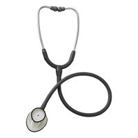
How does a stethoscope work to detect heart and lung sounds?
A stethoscope works by amplifying internal body sounds, allowing healthcare professionals to listen to heart and lung functions. It consists of a chest piece with a diaphragm and/or bell, tubing, and earpieces.
The diaphragm is a flat, circular piece that picks up high-frequency sounds. When placed on the patient's skin, it vibrates in response to sound waves produced by the heart and lungs. These vibrations are transmitted through the stethoscope's tubing to the earpieces, where they are amplified and heard by the user. The diaphragm is particularly effective for detecting high-pitched sounds like normal heartbeats, breath sounds, and certain murmurs.
The bell, on the other hand, is a hollow, cup-shaped piece that is more sensitive to low-frequency sounds. It is used to detect lower-pitched sounds such as heart murmurs and some abnormal lung sounds. When the bell is placed lightly on the skin, it picks up vibrations from the body surface, which are then transmitted through the tubing to the earpieces.
The tubing of the stethoscope is designed to be airtight to prevent sound loss and to ensure that the sound waves travel efficiently from the chest piece to the earpieces. The earpieces fit snugly into the user's ears, further isolating the sounds and reducing external noise interference.
By using a stethoscope, healthcare providers can assess the rate, rhythm, and quality of heartbeats, detect abnormal heart sounds like murmurs or gallops, and evaluate lung sounds for wheezes, crackles, or decreased breath sounds, aiding in the diagnosis of various medical conditions.
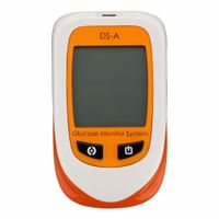
What is the importance of blood glucose monitoring in diabetes management?
Blood glucose monitoring is crucial in diabetes management as it provides real-time data on blood sugar levels, enabling individuals to make informed decisions about their diet, physical activity, and medication. Regular monitoring helps in maintaining blood glucose within the target range, reducing the risk of both short-term complications like hypoglycemia and hyperglycemia, and long-term complications such as neuropathy, retinopathy, and cardiovascular diseases.
By tracking blood glucose levels, individuals can identify patterns and understand how different foods, activities, and stressors affect their blood sugar. This information is vital for adjusting insulin doses or other medications, ensuring optimal glycemic control. It also aids healthcare providers in tailoring treatment plans to the individual's needs, improving overall diabetes management.
Moreover, consistent monitoring empowers individuals with diabetes, fostering a sense of control and responsibility over their health. It encourages proactive management, which can lead to better adherence to treatment regimens and lifestyle modifications. For those using insulin, especially, frequent monitoring is essential to prevent dangerous fluctuations in blood sugar levels.
In addition, blood glucose monitoring is a key component of diabetes education, helping individuals recognize symptoms of abnormal blood sugar levels and take corrective actions promptly. It also facilitates communication with healthcare providers, as accurate records of blood glucose readings can guide clinical decisions and adjustments in therapy.
Overall, blood glucose monitoring is a fundamental aspect of diabetes management, essential for achieving and maintaining good glycemic control, preventing complications, and enhancing the quality of life for individuals with diabetes.
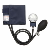
How do blood pressure monitors help in assessing heart health?
Blood pressure monitors are crucial tools in assessing heart health as they provide vital information about the force of blood against the walls of the arteries. Regular monitoring helps in early detection and management of hypertension, a major risk factor for heart disease. By measuring systolic and diastolic pressures, these devices offer insights into the heart's functioning and the condition of the arteries. Elevated readings can indicate increased risk for heart attack, stroke, and other cardiovascular issues. Consistent monitoring allows for tracking changes over time, helping healthcare providers adjust treatment plans and lifestyle recommendations. Additionally, blood pressure monitors can detect irregular heartbeats, which may signal underlying heart conditions. By providing real-time data, they empower individuals to take proactive steps in managing their heart health, ultimately reducing the risk of serious complications.
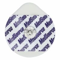
What is the purpose of an ECG and how is it performed?
An electrocardiogram (ECG or EKG) is a medical test used to assess the heart's electrical activity. Its primary purpose is to detect and diagnose various heart conditions, such as arrhythmias, heart attacks, and other cardiac abnormalities. By measuring the timing and strength of electrical signals as they travel through the heart, an ECG can provide valuable information about heart rate, rhythm, and the overall health of the heart muscle.
The procedure for performing an ECG is straightforward and non-invasive. It typically involves the following steps:
1. **Preparation**: The patient is asked to lie down on an examination table. To ensure good electrical contact, areas on the skin where electrodes will be placed are cleaned, and sometimes hair is shaved if necessary.
2. **Electrode Placement**: Small, sticky electrodes are attached to the patient's skin at specific locations, usually on the chest, arms, and legs. A standard ECG uses 10 electrodes to create 12 different views (leads) of the heart's electrical activity.
3. **Recording**: The electrodes are connected to an ECG machine via wires. The machine records the electrical signals from the heart and displays them as waveforms on a monitor or prints them on paper. This process usually takes a few minutes.
4. **Analysis**: A healthcare professional analyzes the waveforms, looking for any irregularities in the heart's rhythm, size, and position of the heart chambers, or signs of damage to the heart muscle.
An ECG is a quick, painless, and safe procedure that provides critical information for diagnosing heart conditions, guiding treatment decisions, and monitoring the effectiveness of interventions.
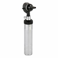
What tools are used in ENT diagnostics and what do they examine?
ENT (Ear, Nose, and Throat) diagnostics utilize a variety of tools to examine and diagnose conditions related to these areas:
1. **Otoscope**: Used to examine the ear canal and eardrum. It helps in diagnosing infections, blockages, and eardrum perforations.
2. **Tuning Fork**: Assesses hearing and bone conduction. It helps in differentiating between conductive and sensorineural hearing loss.
3. **Audiometer**: Measures hearing acuity. It is used in audiometry tests to evaluate hearing loss levels.
4. **Nasal Speculum**: Used to widen the nostrils for examination of the nasal cavity. It helps in identifying obstructions, infections, or structural abnormalities.
5. **Rhinoscope**: A type of endoscope for nasal examination. It provides a detailed view of the nasal passages and sinuses.
6. **Laryngoscope**: Used to examine the larynx and vocal cords. It is essential for diagnosing voice disorders, throat infections, and tumors.
7. **Endoscope**: Flexible or rigid, used for detailed examination of the nasal passages, sinuses, throat, and sometimes the ear. It aids in diagnosing structural issues, infections, and tumors.
8. **Tympanometer**: Assesses the function of the middle ear. It helps in diagnosing fluid in the middle ear, eardrum perforations, and Eustachian tube dysfunction.
9. **Stroboscope**: Used to examine vocal cord vibrations. It is crucial for diagnosing voice disorders and vocal cord lesions.
10. **CT/MRI Scans**: Imaging tools used to get detailed pictures of the sinuses, ear structures, and throat. They help in diagnosing tumors, infections, and structural abnormalities.
11. **Biopsy Tools**: Used to take tissue samples from the ear, nose, or throat for pathological examination, crucial for diagnosing cancers or other tissue abnormalities.
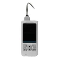
How do pulse oximeters measure oxygen saturation and heart rate?
Pulse oximeters measure oxygen saturation (SpO2) and heart rate using a non-invasive method called photoplethysmography. The device typically consists of a small probe that is clipped onto a thin part of the patient's body, such as a fingertip or earlobe.
The probe contains two light-emitting diodes (LEDs) that emit light at two different wavelengths: red (around 660 nm) and infrared (around 940 nm). These wavelengths are chosen because oxygenated and deoxygenated hemoglobin absorb light differently at these wavelengths. Oxygenated hemoglobin absorbs more infrared light and allows more red light to pass through, while deoxygenated hemoglobin absorbs more red light and allows more infrared light to pass through.
As the light passes through the tissue, a photodetector on the opposite side of the probe measures the intensity of light that has passed through. The device calculates the ratio of absorbed red and infrared light, which correlates with the proportion of oxygenated hemoglobin in the blood. This ratio is then used to estimate the percentage of hemoglobin molecules in the blood that are saturated with oxygen, providing the SpO2 reading.
For heart rate measurement, the pulse oximeter detects the pulsatile changes in blood volume with each heartbeat. As the heart pumps blood, the volume of arterial blood in the tissue increases, causing a corresponding increase in light absorption. By analyzing these periodic changes in light absorption, the device can determine the pulse rate.
The pulse oximeter continuously updates these readings, providing real-time monitoring of both oxygen saturation and heart rate. This method is widely used due to its simplicity, speed, and non-invasive nature, making it a valuable tool in medical settings.